Ultrasound of the Gastrointestinal Tract Part I
Course Content
Topic: Ultrasound of the Gastrointestinal Tract Part I
Abdominal ultrasonography (AUS) is frequently used in the diagnostic work-up of dogs with suspected gastrointestinal disorders. Previous reports have described the ultrasonographic appearance of the normal canine gastrointestinal tract, gastrointestinal neoplasia, intestinal foreign bodies, obstruction, enteritis, intussusception and lymphangiectasia. There have been numerous ultrasonographic studies of the intestinal wall, including measurements of intestinal wall thickness. Increased thickness of the intestinal wall and altered echogenicity of wall layers have been reported in some dogs with diarrhoea; however, there was no association between ultrasonographic intestinal wall thickness and either the histological diagnosis or the response to treatment in dogs with diarrhoea. It has been suggested that mucosal echogenicity may be a more accurate indicator of IBD than intestinal wall thickness in dogs with chronic diarrhoea.
-
What is the clinical utility of AUS in dogs with acute versus chronic gastro-intestinal disease?
-
What is contrast enhanced ultrasonography (CEUS) and how does it allow discrimination between IBD affected dogs and healthy dogs by evaluation of time-intensity curves?
-
Does CEUS provide useful information for monitoring therapeutic response in IBD patients?
-
How are the diagnostic quality assessments of abdominal ultrasound examinations influenced by the performing veterinarians with varying levels of expertise?
Course Content
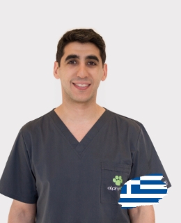
Ioannis Panopoulos
Ioannis graduated from the Veterinary School of the University of Thessaly in 2007.
⦁ In 2010 he completed a two-year training program in pet imaging at the University of Bologna, Italy. During this time he was trained in handling and diagnosis in radiology, ultrasound, computed tomography, magnetic resonance imaging and radioscopy.
⦁ In 2010 he started his doctoral dissertation in the Department of Radiology at the University of Bologna, Italy under the guidance of Professor Mario Cipone and Professor Alessia Diana.
⦁ In 2011 he visited the Department of Radiology of the University of Montreal, Canada for short training under the supervision of Professor Marc-Andre d'Anjou.
⦁ In 2013 he supported his doctoral dissertation on the anatomical study of the anatomical features of the cat's thoracic cavity with axial angiography.
⦁ Since 2011 he has been working at the Alphavet radiodiagnostic center where since 2018 he has established postgraduate positions. www.alphavet.gr
⦁ In 2016 he started further training in radiology and is a trainee of the European College of Radiology (ECVDI). In parallel with his training, he works at the veterinary clinic Istituto Veterinario di Novara in Italy under the supervision of Edoardo Auriemma, a graduate of the European College of Radiology.
⦁ In 2020 he successfully completed the training and examinations of the European College of Radiology (ECVDI) and was sworn in as a Graduate of the European College of Radiology (DipECVDI).
⦁ Since 2020 he has been recognized as a specialized radiologist and is a member of the EBVS.
⦁ Participates as a researcher and speaker in many scientific research programs and conferences in Greece and abroad.
